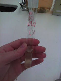
Today in lab, we identified our unknown bacteria (C) is B. cereus.
Bacillus cereus Results of Tests:
Shape: Bacilli
Arrangement: Chained
Spores: Yes
Gram's Stain: Gram Positive
Motility: Motile
Capsules: No capsules
Nutrient Agar: Raised/Cream/Tan colored, Circular with irregular edges
Nutrient Broth: Raised/Cream/Tan colored, Circular with irregular edges
Gelatin Stab: Positive (liquid)
Oxygen Requirements: Facultative Anaerobe
Optimum Temperature: 30 degrees Celsius
Glucose Test: Results were yellow and therefore positive
Lactose: Red and therefore negative
Sucrose: Yellow-Positive-No Gas
Mannitol: Did not grow
Gelatin Liquefaction: Positive, formed liquid
Starch: Positive
Caesin: Positive
Fat: Positive
Indole: Negative
Methyl Red: Negative, high pH
Voges-Proskauer: Negative
Citrate: Negative
Nitrate: Positive
Oxidase: Positive
Litmus Milk Coagulation: Running Curd
We also worked on our yogurt project, and we were successful in growing the different brands of yogurt! Except for Chobani Greek Yogurt- It was the only one that did not grow, so next time, we will thoroughly mix it in with the milk to see if it makes a difference.
Monday, December 12, 2011
Saturday, December 10, 2011
11/22: ELISA Test
Today in lab we did an ELISA test. ELISA stands for Enzyme Linked ImmunoSorbent Assay. It's a test that tests for the Human Immunodeficiency Virus which has antigens and may have antiserum which are antibodies.
11/17: Yogurt and UV light Results
We looked at the yogurt we made on 11/15. We successfully made yogurt! It was white and had a thick consistency. People tasted it and it tasted tart.
Also, we got the results to the UV-C light experiment and did not expect what we found. Our bacteria grew, so the UV light did not kill the bacteria.
Also, we got the results to the UV-C light experiment and did not expect what we found. Our bacteria grew, so the UV light did not kill the bacteria.
11/15: Yogurt Lab and Effect of UV light on Bacterial Growth
In this lab, we made yogurt by doing the following steps:
1. Heated milk in the microwave for 7 to 10 minutes until it boiled
2. Added Probiotic Substances, Kefir, Yogurt with Live and Active Cultures
3. Mix together
4. 40 degree celsius environment
5. Wait for 7-9 hours
(results will be checked on 11/17)
We also tested the effect of UV-C light on bacterial growth. We did this by inoculating a agar plate with our bacteria and then we set the plate under the light for 30 seconds. We expect that it will kill the bacteria, and when we check the results, no bacteria will grow.
Dr. P did another test to check the effect of UV-C light on bacterial growth by putting bacteria in a glass water and put a special UV light pen in the glass (with water and bacteria). The UV light pen kills the bacteria making the water safe to drink.
11/10:Oxidase
Results for tests from 11/8
Citrate: Negative for Citrate
Nitrate: Positive for Nitrate
Indole: Negative for Indole
Urea: Bacteria grew at bottom, and color was yellow which means it does not break down urea.
Antibiotic Test: To determine if the antibiotics kill our bacteria. The measurements determine whether the bacteria is sensitive to the antibiotic.
Antibiotics that break down cell wall
1) Penicillinà 0mm
2) Vancomycinà 20 mm
Nucleic Acid
3) Novabiocinà 29 mm
Protein
4) Tetracyclineà 15 mm
5) Erythromycinà 34 mm
6) Chloramphenitolà 29 mm
7) Neomycinà 20 mm
The next test we did was the oxidase test to determine if our bacteria has cytochrome oxidase, which is an enzyme. When we inoculated it, it turned purple, therefore it his cytochrome oxidase.
Wednesday, November 9, 2011
Lab 11/8: Nitrate, Citrate, Urea, Indole
Tests for today's lab: Nitrate, Citrate, Urea, Indole:
Nitrate Test determines whether or not our bacteria reduces Nitrate to Nitrite
Citrate Test is to see if our bacteria utilizes citrate as it's Carbon Source.
Urea Test is to determine if our bacteria produces the "urease enzyme"
Indole Test is to see if our bacteria can resist the nucleic acid called tryphotan and make what is known as Indole.
We inoculated 4 test tubes for the following tests: Nitrate, Citrate, Urea, and Indole. In order to find the results of urea, we will check it in 24 hours. For Nitrate and Indole, a reagent is added in the next lab on Thursday. We also made a streak plate to test 7 different antibiotics on our bacteria. (Say what happens in each of the tests).
Lab 10/27: Mannitol, MacKonkey, EMB tests
Today we inoculated 3 agar plates, Mannitol, Mackonkey, and EMB. We'll look at the results on Tuesday, and figure out whether our bacteria grows in these conditions or not. All three plates are differential mediums to determine the metabolizing capabilities of our bacteria.
The Mackonkey test is a test to differentiate enteric bacteria.
Mannitol Salt is a test to see if our bacteria can grown in especially salty conditions.
EMB: Eosin Methylene Blue test is to differentiate fecal coliforms.
The Mackonkey test is a test to differentiate enteric bacteria.
Mannitol Salt is a test to see if our bacteria can grown in especially salty conditions.
EMB: Eosin Methylene Blue test is to differentiate fecal coliforms.
Lab 10/25: Water Filtration Plant and tests
We went to the water filtration plant today to observe the process of treating the water of Steubenville. We also started three tests,
Lab 10/20: Glucose, Lactose, Sucrose, TSIA, Gelatin Results!
Glucoseà Yellow, meaning it used the sugar, but there was no gas in it (if there was, bubbles would have appeared)
Lactoseà Red
SucroseàYellow, meaning it used the sugar, there was no gas present here either.
TSIAà Red Slant with a Yellow Butt. The yellow butt indicates that the bacteria maintained growth in acidic conditions, but only the bacteria used the glucose.
Gelatinà We noticed that the bacteria grew in this slant after observing it in room temperature, and then we put this slant in the refrigerator for 15 minute. After taking it out, we noticed that it turned to liquid and therefore, the Gelatin test was positive (a negative test would have been solid).
Lab 10/18: Sugars, Litmus and TSIA tests
Today, we inoculated 6 different tubes including: Glucose, Litmus Milk, Lactose, TSIA (Triple Sugar Ion Agar), Sucrose, Gelatin. These tests will show whether or not our bacteria will or will not grow in these conditions.
Lab 10/13: Starch and Caesin Hydrolysis
(Preserving our environmental bacteria)
We found that our bacteria grew in anaerobic conditions, as well as aerobic conditions; therefore, it’s a facultative aerobic bacteria. There was more growth on the top of the Thioglycolate broth, but it still grew throughout the tube. Our next tests are the Starch Hydrolysis Test and the Caesin Hydrolysis Test. We inoculated a Starch Agar plate and another plate for Casein. We also took our environmental bacteria (collected from the bathroom door handle) and put it in a container, where it will be preserved.
Lab 10/11: Environmental Conditions
Thioglycollate Broth (Above)
Today, we tested our bacteria to figure out whether it uses oxygen, the amount it uses, and if it is tolerant of oxygen. We did this in 2 ways: 1) by inoculating a thioglycollate broth tube, which shows the amount of Oxygen bacteria uses. 2) by putting a sample of our bacteria in the GasPak which makes the environment completely free of O2. In the GasPak, there's an envelope with a paper that indicates whether oxygen is present in the container, or if the container is free of oxygen. You use catalyst called Palladium, which takes out CO2 and replaces it with O2. If there’s O2, the paper turns blue, if there’s no O2, the paper is white. This is how you can tell whether the GasPak is free of O2 or not. We also did the candle in the jar experiment. The burning candle is fueled by the O2 in the environment, but once the cap is put on the jar, all of the oxygen in the jar is used up by the flame and the environment in the jar is free of O2, and CO2 is abundant. Therefore, the flame’s fuel runs out and it dies down.Saturday, October 22, 2011
Lab 9/29: Capsule Staining
Today, we determined whether our Unknown Bacteria C has a capsule around it or not. Capsules are highly organized firmly attached polysaccharide accessories to the cell wall which may contain lipids or proteins. If our bacteria is encapsulated, it is able to protect itself from being phagocytized or ingested. Therefore, encapsulated bacteria are able to survive longer in the body than non encapsulated bacteria.
The first picture shows the stains that we used: Nigrosin Stain, and Safranin
The Nigrosin was a black stain that we smeared across the slide with our bacteria on it. Its the stain that brings out the actual capsule. The Safranin stain stained our bacteria.
Encapsulated Bacteria (note: Not our unknown, just an example of what Encapsulated bacteria would look like)
The next picture reveals our bacteria to be non-encapsulated. Our bacteria has a dark inside and light outside where encapsulated bacteria would have a light inside and dark outside (making it look like white holes in a pink background). The picture directly above is encapsulated.
Lab 9/27: Gram Staining
We determined whether our Unknown Bacteria C was Gram Positive or Gram Negative. In order to do this we:
1. Fixed a slide with a smear of bacteria
2. Stained the fixed slide with Crystal Violet Dye
3. Added Gram's iodine
4. 95% Ethanol was added as a de-colorizer solution which removed some of the stain
5. Added Safranin stain
Above are the fixed slides of our environmental bacteria (from the bathroom door) and our unknown bacteria.
The picture above is our unknown bacteria C
Above is the Gram stain of our Environmental Bacteria
Once the slide was stained, we looked at our bacteria using the oil immersion objective of the Compound Microscope, and determined that our bacteria is Gram Positive.
Lab 9/22: Bacteria Motility
In this class, we tested our unknown C bacteria’s motility. In order to test this, we used a .4% agar tube, and put a sample of our bacteria on a loop and stabbed it straight down into the agar. The .4% agar is solid enough that the bacteria will not spread around, but fluid enough that the bacteria is able to swim, if it is motile. If the bacteria is able to swim, the .4% agar tube will appear cloudy after the bacteria is grown. If it is non-motile, the bacteria will only grow in the stab line, and will not be cloudy.
Results: Unkown Sample C is Motile. Our environmental sample is nonmotile.
Lab 9/20: Determined Shape and Color of Bacteria
In lab, we
1. Inoculated a broth tube with unknown sample C
2. Prepared a Streak plate for the unknown sample C to start colony morphology.
3. Repeated a simple stain for our environmental and unknown sample C.
a. There are 2 types of stains:
i. Acidic, which binds to proteins that are positively chargedà Anionic
ii. Basic, which binds to negatively charged nucleic acid and cellular components à Cationic
4. We determined the shape of our unknown sample C and found that it was:
a. Cream or tan colored
b. Circular shape
c. Had Irregular edges
Wednesday, September 14, 2011
Lab 9/13: Isolation of Bacteria Part III
This week, we took a sample of our bacteria and did 2 things:
1) We made 2 stained slides by putting the bacteria on the slides and dropping 2 different color stains on it (Crystal Violet and Saffron).
2) We got an unknown sample (Sample C) and prepared an Agar Slant. The test tube said the bacteria should be incubated in 30 degrees Celsius, so we put it in the 30 degree Celsius incubator and we'll take it out on Thursday, The color of our sample was whitish yellowish.
For both parts of this lab, we used the Aseptic Technique by sterilizing the loop every time we used it to transfer the bacteria to different petri dishes or containers, and we flamed the test tubes when transferring the unknown bacteria from the original test tube to the two others.
For both parts of this lab, we used the Aseptic Technique by sterilizing the loop every time we used it to transfer the bacteria to different petri dishes or containers, and we flamed the test tubes when transferring the unknown bacteria from the original test tube to the two others.
Subscribe to:
Posts (Atom)


